



Bone Transport

Home / Treatments / Bone Transport

Bone Transport Surgery in India | Ilizarov Technique | Dr. Divya Ahuja

Bone loss is one of the most difficult problems in orthopaedic surgery. Whether it results from a severe road accident, a deep bone infection, a tumour removal, or a surgery that did not heal correctly, a large gap in the bone cannot repair itself without help. Bone transport is the procedure that fills that gap, not with borrowed material from elsewhere in the body, but with brand-new bone that your own body grows during treatment.

Diagram of an internal lengthening nail inserted through the tibia with distraction site marked

What is Bone Transport?

Bone transport is a surgical process where a segment of healthy bone is gradually moved through a defect in the same bone. As that segment moves, new bone forms in the space it leaves behind. This newly formed bone is called the regenerate. Over weeks and months, the regenerate matures into solid, permanent bone that fills the entire defect.

How Does It Work?

The Ilizarov circular fixator is the external frame that makes bone transport possible. It is connected to the bone through fine wires and pins that pass through the skin, and it holds the bone segments in precise alignment throughout treatment.

Key features of the Ilizarov frame in bone transport:

  • Attached externally, so it does not interfere with the zone of injury or infection
  • Allows controlled movement of the bone segment at a rate of 1 mm per day
  • Can be adjusted to correct angular or rotational deformity simultaneously
  • Supports partial weight-bearing from an early stage of recovery
  • Removed once X-rays confirm the bone has fully consolidated

Dr. Ahuja also uses modern hybrid fixation systems that combine an external Ilizarov frame with internal support when appropriate, reducing the overall time the frame must be worn.

Who Needs Bone Transport Surgery?

Bone transport is recommended when a segmental bone defect is present and conventional treatments such as bone grafting or internal fixation alone cannot reliably bridge the gap. The standard threshold is a defect greater than 3 cm in the tibia or greater than 5 cm in the femur, though each case is assessed individually.

Common Conditions That Lead to Bone Transport

Patients referred to Dr. Ahuja for bone transport typically present with one of the following:

  • Severe trauma with bone loss – open fractures where bone was lost or destroyed at the time of injury, most commonly in road traffic accidents
  • Chronic osteomyelitis – long-standing bone infection that required surgical removal of the infected segment, leaving a structural gap
  • Post-tumour resection defects – gaps left after a bone tumour has been excised, where limb salvage is the goal rather than amputation
  • Non-union after prior surgery – bone that has failed to heal after internal fixation, plating, or earlier bone grafting
  • Congenital bone gaps – children and young adults born with missing or underdeveloped bone segments in a limb
  • Failed earlier bone transport – revision cases where the first attempt was incomplete, the frame was removed too early, or the docking site did not fuse

If you have been told your case is too complex for standard orthopaedic treatment, or if you have had a prior surgery that did not work, bone transport is most likely still an option.

Types of Bone Transport Dr. Ahuja Performs

The surgical approach is matched to the size, location, and complexity of the defect. Dr. Ahuja selects from the following techniques based on each patient’s individual assessment.

Monofocal, Bifocal, and Trifocal Bone Transport

Monofocal bone transport uses a single osteotomy and one moving segment. It is the standard approach for defects up to approximately 8 cm. Treatment time is longer because one segment is responsible for closing the entire gap.

Bifocal bone transport uses two osteotomies, creating two segments that move toward each other simultaneously. This is preferred for defects between 8 and 14 cm because it roughly halves the active treatment time compared to monofocal transport.

Trifocal bone transport uses three osteotomies and three active transport zones. It is reserved for very large defects, typically greater than 14 cm, where single or double transport would result in an impractically long treatment duration.

In addition to these standard approaches, Dr. Ahuja offers:

  • Hybrid bone transport combining external Ilizarov fixation with an intramedullary nail, which reduces the time the frame must be worn
  • Acute shortening followed by transport is used when soft tissue coverage is also required
  • Open bone transport for cases with significant overlying wound management needs

The Bone Transport Procedure: What Happens at Each Stage

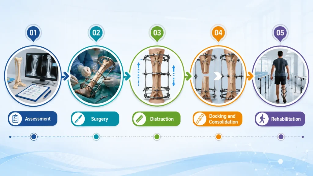
Complete journey of bone transport treatment from assessment to rehabilitation

Bone transport is not a single operation with a short recovery. It is a multi-phase process that unfolds over several months, with the patient playing an active role in daily adjustments. Understanding each stage removes the uncertainty and helps patients commit to the process with confidence.

Stage 1: Preoperative Assessment and Surgical Planning

Before any procedure begins, Dr. Ahuja conducts a thorough assessment using the following:

  • Weight-bearing X-rays to measure the defect length and assess alignment
  • CT scan for three-dimensional bone mapping and osteotomy planning
  • MRI where soft tissue involvement or residual infection needs evaluation
  • Blood markers including ESR and CRP to confirm infection status
  • Digital frame pre-assembly planning so the correct configuration is ready before surgery

In cases where active infection is still present, a staged approach is used. The infected bone and tissue are debrided first, antibiotics are given to achieve infection control, and bone transport begins only once inflammatory markers have settled.

Stage 2: Surgery and Frame Application

The surgical procedure involves the following steps in sequence:

  • A controlled bone cut (osteotomy) is made at a healthy part of the bone, away from the defect
  • This creates the transport segment, a small disc of bone that will be moved through the gap
  • The Ilizarov frame or hybrid fixator is applied externally and connected to the bone with wires and pins
  • Wound closure is performed and the patient is moved to recovery

The surgery itself typically takes two to three hours. Most patients are encouraged to sit up and begin gentle movement of the limb on the same day.

Stage 3: Distraction Phase

After a latency period of approximately seven days, the distraction phase begins. During this phase:

  • The patient turns the adjustment rods on the frame four times daily at evenly spaced intervals
  • Each turn moves the bone segment by 0.25 mm, totalling 1 mm of movement per day
  • New bone (the regenerate) forms in the gap behind the moving segment
  • X-rays are taken every two to three weeks to monitor regeneration quality and alignment
  • Physiotherapy continues throughout to protect joint mobility

The distraction phase lasts approximately one month per centimetre of defect. A five-centimetre defect, therefore, requires around five months of active distraction.

Stage 4: Docking and Consolidation

When the transport segment reaches the far end of the defect and contacts the native bone, this is called docking. From this point:

  • The frame is locked to hold the position while the docking site fuses
  • The regenerate behind the segment continues to mature and harden
  • X-rays at regular intervals confirm the density and structure of the new bone
  • In a minority of cases, a targeted bone graft at the docking site accelerates union
  • The frame is removed once consolidation is confirmed on imaging

Stage 5: Rehabilitation and Return to Activity

Frame removal marks the beginning of the rehabilitation phase:

  • Graduated weight-bearing begins under physiotherapy supervision
  • Muscle strength and joint range of motion are progressively restored
  • Most patients return to unrestricted daily activity within three to four months of frame removal
  • Children typically complete rehabilitation faster than adults
  • Long-term follow-up continues to monitor the regeneration and ensure full bone maturity

Benefits of Bone Transport Over Other Treatments

Patients and referring surgeons frequently ask why bone transport is preferred over bone grafting, the Masquelet technique, or vascularised fibular transfer. The answer depends on the defect size, the presence of infection, and the overall treatment goals.

When Bone Transport May Be Considered

Bone transport offers several advantages that become decisive in the right clinical situation:

  • No donor site required – bone is generated naturally at the defect site, so there is no second surgical wound and no harvest site pain or complications
  • Works in infected cases – the frame is positioned away from the zone of infection, allowing safe reconstruction even after osteomyelitis debridement
  • Corrects multiple problems simultaneously – bone loss, limb shortening, and angular deformity can all be addressed in a single course of treatment
  • Carries healthy tissue forward – as the segment moves, surrounding blood vessels, nerves, and soft tissue advance with it, improving vascularity across the defect zone
  • Highly reliable for large defects – defects greater than 5 cm are poorly managed by bone grafting alone; bone transport has a proven track record at these sizes
  • Limb salvage in extreme cases – patients who would otherwise face amputation have retained their limb through bone transport, including those with defects greater than 15 cm

Treatment Timeline: What to Realistically Expect

One of the most important things to understand before beginning bone transport is the duration of the process and what daily life looks like during treatment.

A Typical Month-by-Month Overview

Phase

Duration

What Happens

Surgery and latency

Week 1

Frame applied, bone cut made, healing begins

Distraction

1 month per cm of defect

Daily frame adjustments, fortnightly X-rays, physiotherapy

Docking and early consolidation

1 to 2 months

Segment contacts native bone, frame locked

Consolidation

2 to 4 months

New bone matures; frame removed when confirmed

Rehabilitation

3 to 4 months post-frame

Weight-bearing progresses, full activity restored

For a 5 cm defect, the total treatment period from surgery to discharge from active follow-up is typically 12 to 15 months. For a 10 cm defect, this extends to approximately 18 to 24 months. Children complete treatment faster at every stage.

What Published Studies Report

Bone transport through the Ilizarov technique achieves bone union rates above 90% in experienced hands. This figure holds across the full range of cases, including large post-infective defects, failed prior surgeries, and defects in both upper and lower limbs.

Possible Outcomes

Patients treated by Dr. Divya Ahuja have:

  • Returned to walking without support or walking aids
  • Resumed physically demanding occupations, including construction, farming, and manual labour.
  • Avoided amputation in cases involving defects greater than 12 cm after severe trauma
  • Can be used as part of treatment for chronic osteomyelitis
  • Regained normal or near-normal limb length and alignment
  • Reported satisfaction with the cosmetic result, as large skin incisions are not required

Dr. Ahuja’s practice includes tibial and femoral defects, forearm bone loss, infected non-unions, and complex revision cases referred from other centres across India.

About the Treating Surgeon

Bone transport is a procedure where the surgeon’s experience with external fixation management determines the outcome more than any other single factor. It is not a procedure that can be effectively performed occasionally by a general orthopaedic surgeon.

Training, Focus, and Clinical Infrastructure

  • Exclusive specialist focus: Dr. Ahuja’s practice is dedicated to limb reconstruction, external fixation, and deformity correction; bone transport is not an occasional procedure here but a clinical core competency
  • Advanced surgical planning: digital tools are used for defect measurement, osteotomy site selection, and frame pre-assembly before every case
  • Inpatient care, imaging and physiotherapy at the treating hospital: procedures are performed at hospitals with modern imaging, intraoperative fluoroscopy, and dedicated physiotherapy departments
  • Remote follow-up support: patients from other cities and abroad receive structured protocols for home frame adjustments and X-ray review between in-person visits
  • Revision case experience: Dr. Ahuja accepts complex referrals from other centres where prior bone transport was incomplete, or complications occurred
  • Transparent consultation process: detailed cost estimates, realistic timelines, and honest discussion of risks are provided before any commitment is made

Frequently Asked Questions about Bone Transport

A surgical procedure that moves a bone segment through a defect, stimulating new bone growth to fill the gap naturally using the Ilizarov technique.

Roughly one month per centimetre of defect, plus two to four months of consolidation. A five-centimetre defect takes around twelve to fifteen months total.

Discomfort is expected but manageable with oral medication. Most patients describe the daily adjustments as mild pressure, not sharp pain.

Yes. Dr. Ahuja uses a staged approach: debridement first, then transport once infection markers settle. The Ilizarov frame stays away from the infected zone.

Turn the frame adjustment rods four times daily, clean pin sites, attend physiotherapy, and come in for fortnightly X-ray reviews.

Above 90% union rate in experienced hands, including in complex post-infective and revision cases.

Pin site infection, joint stiffness, and docking site non-union. All are manageable with proper monitoring, physio, and early intervention.

Costs vary; an estimate is given after assessment. An itemised estimate is provided after the initial consultation and imaging review.

Yes. Children respond exceptionally well. New bone forms faster, treatment is shorter, and growth plates are protected during planning and surgery.

Yes. Revision bone transport is possible when the first attempt was incomplete, the frame was removed early, or the docking site did not fuse.


Diagram of an internal lengthening nail inserted through the tibia with distraction site marked