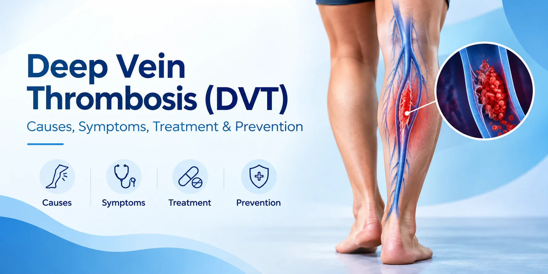
Deep vein thrombosis (DVT) is a blood clot that forms in one of the body’s deep veins, most often in the calf, thigh, or pelvis. It is a serious complication because a fragment can travel to the lungs and cause a pulmonary embolism, which can be fatal. The two biggest risk factors are lower-limb fractures and hip, knee, or leg surgery, along with prolonged immobilisation, cancer, obesity, smoking, and inherited clotting disorders. Symptoms include one-sided leg swelling, pain, warmth, and redness, though nearly half of cases have no symptoms until a pulmonary embolism occurs. A duplex ultrasound of the leg confirms the diagnosis. Treatment involves anticoagulant medication for three to six months, compression stockings, and, in selected cases, thrombolysis or an IVC filter. Prevention after orthopedic surgery includes early mobilisation, compression devices, and prescribed blood thinners for up to three months.
What is Deep Vein Thrombosis (DVT)?
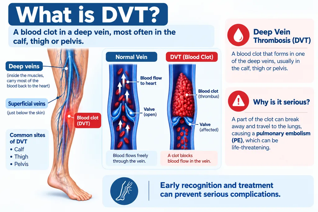
The body has two types of veins in the legs. Superficial veins lie just below the skin, while deep veins run inside the muscles and carry most of the blood back to the heart. A deep vein thrombosis is a blood clot (thrombus) that forms inside one of these deep veins, usually in the calf, thigh, or pelvis.
Once a clot forms, it slows the return of blood to the heart, causing swelling and pain in the affected leg. More seriously, a piece of the clot can break off and travel through the bloodstream, lodging in the lungs. This is called a pulmonary embolism (PE), and it can be fatal if not treated urgently. DVT and PE together form a condition doctors call venous thromboembolism (VTE).
Why DVT Matters in Orthopedic Practice
Two of the strongest risk factors for DVT are directly relevant to orthopedic care:
- A significant injury to the lower body, such as a hip, thigh, or leg fracture
- Surgery involving the hip, knee, or leg, including joint replacement, complex fracture treatment, and Ilizarov hip reconstruction
Prolonged immobilization in a cast, external frame, or bed after major surgery further slows blood flow in the deep veins. Procedures such as limb lengthening may require several months of protected weight-bearing, and each of these situations increases the baseline risk of clot formation.
This is why every well-run orthopedic practice builds DVT prevention into the treatment plan from day one, and why patients should understand what the condition is and how to help protect themselves.
Causes of DVT
Doctors describe three factors, known as Virchow’s triad, that come together to form a deep vein clot.
Slow Blood Flow (Stasis)
When blood does not move freely through the veins, it fails to mix with natural anti-clotting agents and is more likely to clot. Situations that slow blood flow include:
- Bed rest after major surgery or illness
- Immobilization in a cast, brace, or external fixator
- Long-haul flights or bus and train journeys
- Prolonged inability to walk after a fracture
Hypercoagulability
The blood itself may become more prone to clotting. This can happen due to:
- Release of tissue debris, collagen, or fat during surgery
- Bone preparation during hip or knee replacement
- Inherited clotting disorders such as Factor V Leiden
- Cancer, dehydration, and certain medications
- Pregnancy, oral contraceptives, and hormone therapy
Damage to Vein Walls
During major fractures or surgery, retraction of soft tissues and manipulation of bone can injure nearby veins. Damaged vein walls trigger the clotting cascade, contributing to thrombus formation.
Risk Factors for DVT
You are at higher risk of DVT if you have any of the following:
- Recent major surgery, especially of the hip, knee, or leg
- Recent lower-limb fracture or trauma
- Immobilization in a cast, brace, or fixator
- Age above 60 years
- Personal or family history of DVT or pulmonary embolism
- Cancer or ongoing chemotherapy
- Obesity
- Smoking
- Pregnancy and postpartum period
- Long journeys with prolonged sitting
- Oral contraceptive pills or hormone replacement therapy
- Inherited clotting disorders
- Chronic heart, lung, or inflammatory disease
The more risk factors present, the higher the chance of clot formation, and the more aggressive the prevention plan needs to be.
Symptoms of DVT
DVT often develops quietly, but classic warning signs include:
- Swelling of the calf or thigh that does not improve with elevation
- Pain, tenderness, or a cramp-like ache in the affected leg
- Warmth and redness over the skin
- A visible, distended vein pattern
- A firm, cord-like feeling along the course of the vein
Up to half of DVT cases have no leg symptoms at all, and the first sign may be a pulmonary embolism. Symptoms of pulmonary embolism include:
- Sudden shortness of breath
- Sharp chest pain, often worse with deep breathing
- Rapid heart rate
- Coughing, sometimes with blood
- Feeling faint or losing consciousness
Any of these symptoms after a fracture, orthopedic surgery, or long period of immobility need immediate emergency evaluation.
Complications of DVT
Pulmonary Embolism
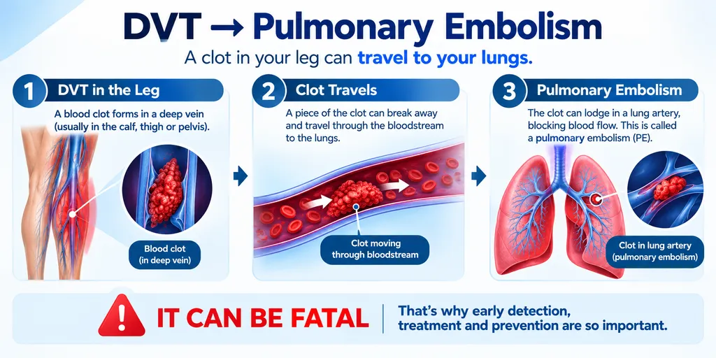
Pulmonary embolism is the most feared complication. When a clot breaks off and blocks blood flow to the lungs, it can cause acute breathlessness, chest pain, low blood pressure, and, in severe cases, death within minutes. It is a medical emergency requiring hospital admission, imaging, and immediate anticoagulation or clot-busting therapy.
Post-Thrombotic Syndrome
Even when the clot itself is treated successfully, damage to the valves inside the vein can cause post-thrombotic syndrome (PTS). Blood pools in the leg, raising venous pressure and leading to chronic swelling, aching, skin discoloration, brownish pigmentation, and, in advanced cases, non-healing venous ulcers around the ankle. PTS can significantly reduce quality of life for years after the initial DVT.
How DVT Is Diagnosed
DVT diagnosis combines clinical assessment with targeted testing.
- Wells score, a clinical scoring system, estimates the probability of DVT before ordering tests
- D-dimer blood test measures a breakdown product of clots. A negative result in a low-risk patient effectively rules out DVT
- Duplex ultrasound is the standard imaging test. It combines traditional ultrasound with Doppler flow to visualise the vein and detect the clot. It is non-invasive and radiation-free
- MRI venography is used occasionally for pelvic and thigh clots that are difficult to see on ultrasound
- CT pulmonary angiogram is used when pulmonary embolism is suspected
- Contrast venography is rarely used today because of its invasive nature
Early diagnosis is critical because untreated DVT is far more dangerous than the medications used to treat it.
Treatment of DVT
The goals of treatment are to stop the clot from growing, prevent it from breaking off, and reduce the risk of another clot forming.
Anticoagulants (Blood Thinners)
Anticoagulants are the mainstay of DVT treatment. Common options include:
- Direct oral anticoagulants (DOACs) such as rivaroxaban, apixaban, and dabigatran, which have simplified DVT care since they do not require regular blood monitoring
- Low molecular weight heparin (LMWH), such as enoxaparin, is given as a subcutaneous injection and is often used to start treatment and in pregnancy
- Unfractionated heparin, given intravenously in hospital
- Warfarin, an older oral agent that needs regular INR monitoring and dietary care
Duration of treatment typically ranges from 3 to 6 months, and longer in patients with recurrent clots or persistent risk factors.
Compression Therapy
Graded elastic compression stockings help reduce leg swelling, prevent pooling of blood, and lower the risk of post-thrombotic syndrome. They are usually worn during the day for months after the acute DVT.
Thrombolytics
Clot-dissolving drugs are reserved for large clots with severe symptoms or when there is a very high risk of pulmonary embolism, and are usually delivered directly into the clot through a catheter.
IVC Filter and Surgery
If a patient cannot take anticoagulants or the clot continues to grow despite treatment, an inferior vena cava (IVC) filter may be placed. This small metal device traps clots before they reach the lungs. Surgical removal of a clot is rare and used only in selected cases.
DVT Prevention After Orthopedic Surgery
The risk of DVT is highest 2 to 10 days after major orthopedic surgery and remains elevated for about 3 months. Prevention protocols are used in every patient, but tailored to the individual’s risk.
Early Mobilization and Physiotherapy
The single most effective prevention is movement. Patients are encouraged to:
- Perform ankle pumps, quadriceps contractions, and leg lifts within hours of surgery
- Sit up and stand with support as early as safely possible
- Walk with crutches, a walker, or physiotherapist assistance from day one when the procedure allows
- Continue prescribed exercises at home
Mechanical Prophylaxis
- Graded compression stockings worn on the legs during the immobile period
- Sequential compression devices (SCDs), boot-like sleeves that pulse pressure on the calves to mimic walking
- Elevation of the leg above heart level when resting
Pharmacological Prophylaxis
- Low molecular weight heparin injections such as enoxaparin, or oral DOACs, started shortly after surgery
- Duration depends on the type of surgery, risk profile, and mobility status
- Every patient undergoing complex fracture surgery, hip reconstruction, or limb lengthening receives a structured DVT prevention plan
When to Seek Emergency Care
Call for emergency medical care immediately if you or a family member experiences:
- Sudden shortness of breath
- Sharp chest pain that worsens with breathing
- Coughing up blood
- Rapid heart rate or fainting
- Rapid worsening leg swelling or discoloration
- A firm, hot, extremely painful calf after surgery or immobility
These may indicate DVT or pulmonary embolism, and time is critical.
DVT Prevention Protocols in Orthopaedic Surgery
Dr. Divya Ahuja, MS Ortho, is a Mumbai-based orthopedic and Ilizarov surgeon with over 15 years of experience in trauma and reconstructive care. His practice at the Center for Limb Lengthening and Reconstruction, Mangal Anand Hospital, Chembur, follows a structured DVT prevention protocol for every major fracture, joint reconstruction, Ilizarov procedure, and limb lengthening case. This includes preoperative risk assessment, mechanical prophylaxis with compression devices, appropriate pharmacological prophylaxis, and early mobilisation guided by physiotherapy. Suspected DVT is investigated urgently with duplex ultrasound and managed in close coordination with vascular medicine specialists. For a preoperative consultation with an experienced orthopedic surgeon in Mumbai, you can book online or visit the clinic at Chembur, Thane, Dadar, Vashi, Navi Mumbai, or Tilak Nagar.
Conclusion
Deep vein thrombosis is a serious but largely preventable complication, especially for anyone recovering from a major fracture or lower-limb surgery. Understanding your personal risk, moving as early as your surgeon allows, wearing compression when advised, taking prescribed blood thinners exactly as directed, and reporting any suspicious leg or chest symptoms immediately can protect you from most complications. For personalised DVT risk assessment and comprehensive prevention planning around fracture surgery, Ilizarov reconstruction, or limb lengthening, consult Dr. Divya Ahuja for an evidence-based orthopedic evaluation.
Frequently Asked Questions (FAQs)
What is deep vein thrombosis (DVT)?
Deep vein thrombosis is a blood clot that forms inside one of the deep veins of the body, most commonly in the calf, thigh, or pelvis. It slows blood return to the heart and can be dangerous if a fragment reaches the lungs.
What are the early symptoms of DVT in the leg?
Early symptoms include one-sided calf or thigh swelling, pain or tenderness, warmth, redness, and a firm cord-like feeling along a vein. Many DVTs, however, have no symptoms at all, especially in the first few days after surgery or immobilisation.
What causes DVT?
DVT is caused by three factors together, known as Virchow's triad: slow blood flow from immobilisation, increased blood clotting from surgery or medical conditions, and damage to vein walls during trauma or operation. Cancer, obesity, pregnancy, and smoking add to the risk.
Who is at highest risk of DVT after orthopedic surgery?
Patients undergoing hip and knee replacement, complex fracture surgery, Ilizarov reconstruction, or limb lengthening are at highest risk, especially if they are over 60, obese, smokers, have cancer, or have a personal or family history of DVT.
How is DVT diagnosed?
DVT is diagnosed using a combination of clinical assessment, the D-dimer blood test, and duplex ultrasound of the affected leg. Duplex ultrasound is the most common and reliable test, and is painless, radiation-free, and can be repeated safely.
Can DVT go away without treatment?
Small isolated calf clots occasionally resolve with monitoring, but most DVTs need treatment. Without anticoagulation, clots can grow, break off, and cause pulmonary embolism, or damage vein valves and cause long-term post-thrombotic syndrome with chronic leg swelling and pain.
What is the main treatment for DVT?
The main treatment is anticoagulation with medicines such as rivaroxaban, apixaban, low molecular weight heparin, or warfarin, usually for three to six months. Compression stockings, thrombolysis, and IVC filters are used in selected cases based on clot size and risk.
How can I prevent DVT after surgery?
DVT is prevented after surgery by early movement, ankle-pump exercises, walking with support as soon as safe, wearing compression stockings, using pneumatic compression devices, and taking prescribed anticoagulant medication for the duration advised by your orthopedic surgeon.
What is a pulmonary embolism?
A pulmonary embolism happens when a clot from a deep vein breaks off and blocks blood vessels in the lungs. It causes sudden breathlessness, chest pain, rapid heart rate, and can be fatal. It is a medical emergency needing immediate hospital treatment.
How long am I at risk of DVT after orthopedic surgery?
Risk is highest between two and ten days after major orthopedic surgery and remains elevated for around three months. Patients continue prescribed prophylaxis, mobilisation, and compression therapy well after discharge to protect them during this vulnerable period.