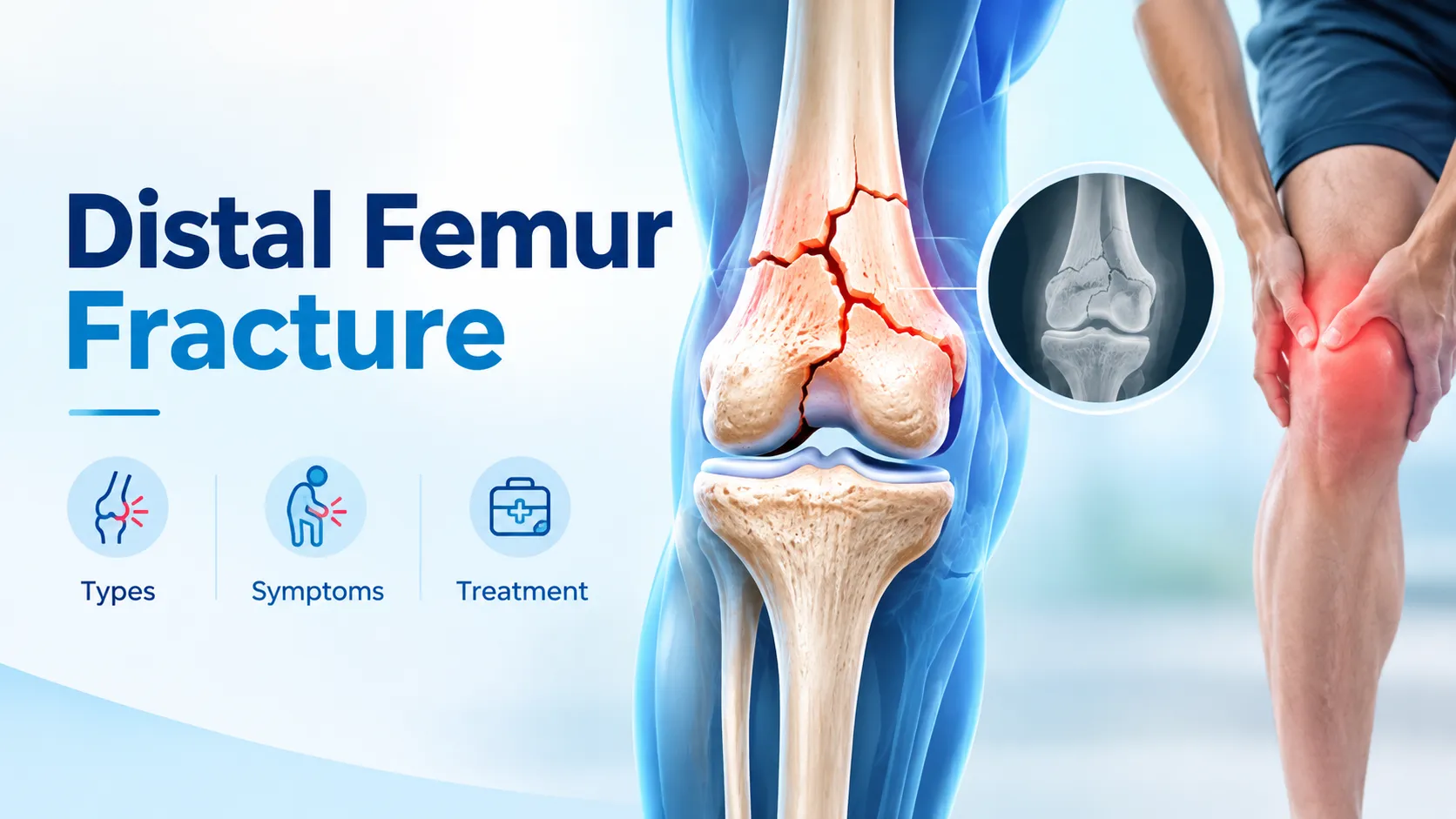
A distal femur fracture is a break in the lower end of the thigh bone, just above the knee joint. It commonly results from high-energy trauma such as road accidents and falls from height in younger adults, and from low-energy falls in elderly patients with osteoporosis. Fractures may be extra-articular (supracondylar), intra-articular (intercondylar or unicondylar), and closed or open. Symptoms include severe knee pain, marked swelling, deformity, and inability to bear weight. Diagnosis relies on X-rays and CT scans, with MRI when soft tissue injury is suspected. Clinicians may treat undisplaced fractures with a cast or brace. Still, most displaced or comminuted fractures need surgical fixation using locking plates, retrograde nails, or Ilizarov and hexapod external frames for complex, open, or infected patterns. Recovery typically takes six to twelve months, and long-term issues include stiffness, arthritis, malunion, and nonunion.
Anatomy of the Distal Femur
The femur, or thigh bone, is the longest and strongest bone in the human body. Its lower end broadens into two rounded prominences called the medial and lateral femoral condyles, which glide against the tibia and patella to form the knee joint.
Key features of this region include:
- Femoral condyles, covered with smooth articular cartilage that allows painless knee motion
- Intercondylar notch, a groove between the condyles that houses the cruciate ligaments
- Supracondylar area, just above the condyles, made of softer cancellous bone that is prone to comminution in injury
- Metaphyseal-diaphyseal junction, where the wider condyles meet the narrower shaft
Because this area is packed with joint surface, ligament attachments, and softer bone, a fracture here can affect knee alignment, motion, and stability all at once, which is why treatment planning is so critical.
What is a Distal Femur Fracture?
A distal femur fracture is a break in the lower part of the thigh bone. Depending on the direction and force of injury, the bone may:
- Crack in a single line above the condyles (supracondylar fracture)
- Split between the condyles into a T or Y shape (intercondylar fracture)
- Involve only one condyle (unicondylar fracture)
- Shatter into multiple fragments (comminuted fracture)
- Break through the skin (open or compound fracture)
Because the knee joint surface is often involved, even a small step-off can lead to pain, stiffness, instability, and post-traumatic arthritis if not restored to normal alignment. Injuries to nearby blood vessels, nerves, ligaments, and the patella are also common in high-energy cases and must be evaluated systematically.
Types of Distal Femur Fractures

Orthopedic surgeons classify these fractures in several ways to plan treatment.
Extra-articular Fractures
These break above the joint surface and do not enter the knee. Supracondylar fractures fall into this group and typically involve the metaphyseal region just above the condyles.
Intra-articular Fractures
These fractures extend into the knee joint. Restoring the cartilage surface to smooth alignment is critical to prevent early arthritis. Intercondylar (T or Y shaped), unicondylar, and coronal (Hoffa) fractures fall in this category.
Open (Compound) vs Closed Fractures
- Closed fractures have intact skin over the break
- Open fractures have bone exposed through the skin, carrying a high risk of infection and always requiring urgent surgical washout
Surgeons commonly use the AO/OTA classification to group distal femur fractures from Type A (extra-articular) through Type C (complete articular with metaphyseal involvement). Higher grades indicate more complex injuries that often need advanced fixation, sometimes with Ilizarov or hybrid external fixators.
Causes of Distal Femur Fractures
The pattern of injury usually depends on the patient’s age and bone quality.
- High-energy trauma in younger adults, including road traffic accidents (especially two-wheeler crashes), falls from height, and industrial injuries
- Low-energy falls in elderly patients with osteoporosis, where a simple stumble at home fractures the weakened bone
- Periprosthetic fractures around a previous knee replacement, a growing problem as more Indians undergo total knee arthroplasty
- Sports injuries with rotational force, particularly in football, kabaddi, and skiing
- Pathological fractures through bone weakened by tumour, cyst, or infection
Symptoms and Warning Signs
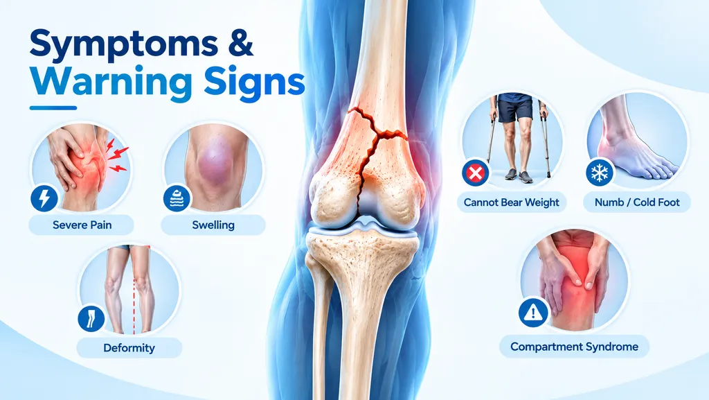
A distal femur fracture usually announces itself immediately after significant trauma. Typical symptoms include:
- Severe pain in the lower thigh and knee that worsens on movement
- Marked swelling and bruising above the knee
- Visible deformity or shortening of the leg
- Inability to bear weight or straighten the leg
- Grinding sensation on any attempt to move
- Pale, cold, or numb foot, which suggests possible vascular or nerve injury
- Tense, hard swelling of the thigh, which is a warning sign of compartment syndrome
Any of these features after trauma calls for immediate emergency evaluation at a hospital equipped for orthopedic care.
Comparison Table: Distal Femur Fracture Patterns and Typical Treatment
| Fracture Pattern | Common Cause | Preferred Treatment |
|---|---|---|
| Undisplaced supracondylar | Low-energy fall, osteoporosis | Cast, brace, protected weight-bearing |
| Displaced supracondylar | High-energy trauma | Locking plate or retrograde nail |
| Intercondylar T or Y | Road accident, fall from height | Articular reduction, plate fixation |
| Unicondylar | Twist, direct impact | Screw or plate fixation |
| Comminuted distal femur | Two-wheeler accident, industrial injury | Locking plate, Ilizarov, hybrid fixation |
| Open comminuted | Road accident, crush injury | Ilizarov or hexapod external fixation |
| Periprosthetic | Fall around a knee replacement | Locking plate or revision arthroplasty |
How a Distal Femur Fracture Is Diagnosed
Diagnosis combines clinical examination with focused imaging. The doctor checks the skin for open wounds, assesses pulses and sensation in the foot, and looks for signs of ligament or nerve injury.
Imaging usually includes:
- X-rays of the knee and full femur in multiple views to identify the fracture and any displacement
- CT scan to map the fracture pattern in three dimensions, essential for planning surgery in intra-articular fractures
- MRI scan when soft tissue or ligament injury is suspected
- CT angiography or Doppler if vascular injury is suspected
A CT scan is particularly valuable for comminuted and intercondylar patterns because it reveals hidden joint fragments that plain X-rays may miss.
Emergency Care for Complex Distal Femur Fractures
Some distal femur fractures need urgent hospital care before any definitive surgery.
Open fractures require surgical washout and debridement within a few hours to reduce the risk of deep bone infection. When infection does develop, specialised bone infection treatment is essential to salvage the limb.
Severe thigh swelling can raise pressure inside the muscle compartments and cut off blood supply. This is a limb-threatening emergency requiring immediate fasciotomy, where the surgeon releases the tight muscle sheaths through vertical incisions.
When the skin and soft tissues are too damaged for immediate plating, a temporary knee-spanning external fixator is applied to maintain alignment for one to two weeks. Definitive fixation follows once the swelling and soft tissue settle. This staged strategy is a cornerstone of modern management for high-energy distal femur injuries.
Definitive Treatment of Distal Femur Fractures
Nonsurgical Treatment
Nonsurgical care is limited to undisplaced fractures, minor stress fractures, and elderly patients who cannot tolerate surgery. It involves a long-leg cast or hinged knee brace for six to twelve weeks, strict non-weight-bearing initially, and serial X-rays to check for late displacement.
Surgical Fixation
Most displaced distal femur fractures need surgical fixation to restore alignment and joint congruity. Common techniques include:
- Locking condylar plate, the workhorse implant for most distal femur fractures, offering stable fixation even in osteoporotic bone
- Retrograde intramedullary nail, inserted through the knee, useful for supracondylar patterns without significant joint involvement
- Percutaneous screws for simple articular splits
- Bone grafting to fill metaphyseal defects in comminuted patterns
- Distal femoral replacement in elderly patients with severe articular comminution or periprosthetic fractures
Ilizarov and Other Reconstruction Methods
In highly comminuted, open, or infected distal femur fractures, and in cases where plating is unsafe due to poor soft tissue, Ilizarov ring fixation, hexapod frames such as Ortho-SUV and Hexapod, and hybrid external fixators offer a powerful alternative. These frames allow:
- Stable fixation without further stripping of the soft tissues
- Gradual correction of alignment and length
- Early weight-bearing on the injured limb
- Simultaneous management of infection, bone loss, and deformity
For segmental bone loss after severe trauma, bone transport and bone gap reconstruction techniques rebuild the thigh bone gradually, avoiding amputation in most cases.
Recovery and Rehabilitation
Recovery from a distal femur fracture is measured in months, not weeks. Even after successful surgery, weight-bearing is usually restricted for six to twelve weeks to protect the joint surface while it heals. Rehabilitation typically follows three phases:
- Phase 1 (0 to 6 weeks): Pain control, gentle knee range-of-motion, and non-weight-bearing mobilisation with crutches or a walker
- Phase 2 (6 to 12 weeks): Partial and progressive weight-bearing, strengthening of the quadriceps and hamstrings, and gait retraining
- Phase 3 (3 to 6 months): Full weight-bearing, functional retraining, and gradual return to work and low-impact activity
Return to running or contact sports typically takes six to twelve months, depending on the fracture pattern, quality of reduction, and rehabilitation compliance.
Long-Term Complications
Even well-treated distal femur fractures can have long-term consequences, especially when the joint surface was severely damaged. Common issues include:
- Knee stiffness and loss of motion, particularly loss of full flexion
- Post-traumatic osteoarthritis of the knee
- Malunion with the leg healing in a bowed, twisted, or shortened position
- Nonunion of a fracture, where the bone fails to heal despite adequate time and fixation
- Bone loss or deep infection after open fractures
- Chronic pain and instability
- Implant failure or breakage in inadequately protected fractures
Post-traumatic limb length discrepancy, angular deformity, and hardware failure often need dedicated reconstructive surgery, and early evaluation makes a substantial difference to long-term outcomes.
Why Choose Dr. Divya Ahuja for a Distal Femur Fracture
Dr. Divya Ahuja, MS Ortho, is a Mumbai-based orthopedic and Ilizarov surgeon with over 15 years of experience in complex trauma and limb reconstruction. His practice at the Center for Limb Lengthening and Reconstruction, Mangal Anand Hospital, Chembur, is specifically set up for distal femur and periarticular knee fractures, including:
- Acute complex distal femur fractures needing plating or Ilizarov fixation
- Open and infected fractures with soft tissue loss
- Nonunion and malunion after previous fracture surgery
- Post-traumatic knee malalignment and stiffness
- Bone gap and segmental defects treated with bone transport
He uses software-assisted planning with TraumaCad and modern six-axis correction systems including Ortho-SUV, Hexapod, and Deftfix. Consultation addresses, days and timings are listed on the Contact page. To book a consultation with an experienced orthopedic surgeon in Mumbai, you can book online or visit the clinic.
Conclusion
A distal femur fracture is one of the most demanding injuries in orthopedic surgery because it involves the largest weight-bearing bone in the body and the knee joint at the same time. Prompt diagnosis, careful surgical planning, and expert use of modern fixation techniques from locking plates to Ilizarov reconstruction are the keys to a good long-term outcome. If you or a family member has sustained a distal femur fracture, or you are dealing with nonunion, malalignment, or post-traumatic arthritis after an old injury, consult Dr. Divya Ahuja for a detailed evaluation and evidence-based treatment plan.
Frequently Asked Questions
What is a distal femur fracture?
A distal femur fracture is a break in the lower end of the thigh bone, just above the knee. It may or may not involve the knee joint surface. Many require surgery to restore alignment and prevent long-term stiffness or arthritis.
What causes a distal femur fracture?
High-energy trauma such as road traffic accidents and falls from height in younger adults, and low-energy falls in elderly patients with osteoporosis, are the main causes. Periprosthetic fractures around a previous knee replacement are also increasingly common.
How long does a distal femur fracture take to heal?
Most distal femur fractures take three to six months to unite on X-ray, though full functional recovery typically takes six to twelve months. Complex, open, or infected fractures may take longer, especially when Ilizarov reconstruction or bone transport is required.
Do all distal femur fractures need surgery?
No. Undisplaced fractures and certain elderly patients unfit for surgery can be managed with a cast or brace. However, most displaced fractures, all significant intra-articular fractures, and open fractures require surgical fixation to achieve the best long-term outcome.
Can I walk after a distal femur fracture?
Not immediately. Full weight-bearing is usually delayed for six to twelve weeks to protect the healing bone and joint. Patients start with crutches or a walker, progress to partial weight-bearing on the doctor's advice, and typically resume normal walking around three months.
What is Ilizarov fixation for a distal femur fracture?
Ilizarov fixation uses a circular external frame with fine wires and rings to stabilise the fracture without extensive incisions. It is especially useful for open, infected, comminuted, or high-energy distal femur fractures, allowing bone healing, early weight-bearing, and gradual deformity correction.
What is compartment syndrome after a femur fracture?
Compartment syndrome is severe pressure build-up inside the thigh muscles that cuts off blood supply. It causes intense pain, tight swelling, and numbness. It is a surgical emergency requiring urgent fasciotomy within hours to save the muscles and nerves of the leg.
What are the complications of a distal femur fracture?
Common complications include knee stiffness and loss of motion, post-traumatic arthritis, malunion with limb bowing or shortening, nonunion, deep bone infection, chronic pain, and implant failure. Long-term outcomes depend on fracture severity, quality of reduction, and rehabilitation.
Can a distal femur fracture heal without surgery?
Undisplaced fractures in a well-fitting long-leg cast can heal without surgery, particularly in elderly patients who cannot tolerate anaesthesia. Displaced or comminuted fractures usually need internal or external fixation to hold alignment while they unite; otherwise, nonunion may develop.
Where can I get treatment for a complex distal femur fracture in Mumbai?
Dr. Divya Ahuja treats complex distal femur fractures at the Center for Limb Lengthening and Reconstruction, Mangal Anand Hospital, Chembur, using modern locking plates, retrograde nailing, Ilizarov, hexapod, and bone-transport techniques for acute injuries and post-traumatic reconstruction.